Campus Moncloa
Campus of International Excellence

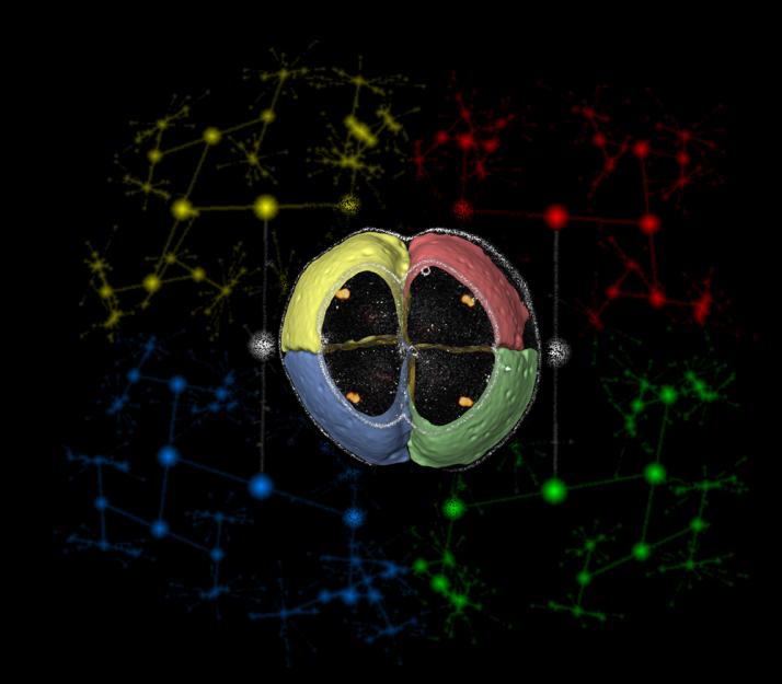
Cartography of a zebrafish embryo (Miguel Luengo Oroz). Award for scientific photography category of i-Health cluster.
Cartography of a zebrafish embryo The image has been obtained by a new microscopy technique that allows to record 3D and high resolution videos on the in-vivo embryo development of a zebrafish without disturbing the process. The central part of the image shows the first 4 cells of a zebrafish embryo in 3D. The four cellular membranes have been automatically colored using mathematical algorithms able to recognize the cell shapes. In addition, the algorithms allow tracking the position of each cell during the embryo development, therefore reconstructing the cell lineage tree and generating the background image that shows the real positions of each cell division from the 4-cell stage to the 1024-cell stage. The circles correspond to locations of cell divisions and the lines to cell displacements. The study of zebrafish embryogenesis has applications in cancer and stem cell research. Luengo-Oroz, M.A., et al. "Methodology for Reconstructing Early Zebrafish Development From In Vivo Multiphoton Microscopy". IEEE Transactions in Image Processing,21(4):2335-2340. Apr.2012 Olivier, N., Luengo-Oroz, M.A., Duloquin, et al. "Cell Lineage Reconstruction of Early Zebrafish Embryos Using Label-Free Nonlinear Microscopy".Science,329(5994):967-971.20 Aug.2010
